

1. Background
Antimicrobial resistance is one of the World Health Organization’s top 10 threats to global health. And unfortunately, it is very much on the rise. The cost of antimicrobial resistance to the economy is substantial. Not only does it lead to death and disability, but lengthy illness results in extended hospital stays. This in turn increases the need for more expensive medicines and financial challenges for those impacted. Without effective antimicrobials, “the success of modern medicine in treating infections, including during major surgery and cancer chemotherapy, would be at increased risk” (WHO, 2020). Before we delve deeper into antimicrobial resistance and optimising nanofiber-based biosensors, let’s take a step back and have a brief look at what sparked this research since this project aims to optimise the initial research.
2. Initial research: Quantification of CD4+ cell count via a nanofiber-based biosensor
Mr Alexander Lloyd, a PhD Candidate in the Department of Electric and Electronic Engineering, Stellenbosch University, conducted research for his postgraduate studies on the quantification of CD4+ cell count via a nanofiber-based biosensor. Antigen-substrate binding has been found to cause a change in the resistance of conductive nanofiber substrates. Alexander’s thesis analysed this phenomenon with a view to produce sensors capable of quantifying CD4+ cell count. Producing nanofiber substrates that are both robust and cost-effective remains a challenge. Alexander explains it as follows:
“Firstly, I wanted the fibres to be nanofibers i.e., their diameter must be sub-micron. The obvious benefit of that is a greater surface area to volume ratio. Secondly, I wanted my nanofibers to be intrinsically conductive. The whole polypyrrole coating process is tricky to reproduce and sort of uneven, plus it’s an additional processing step. So, if I could get rid of that it would be ideal. And then I also wanted my nanofibers to be physically robust.”
Previous methods included coating non-conductive nanofibers with a non-uniform conductive coating or using carbon nanofiber mats that were very susceptible to mechanical stress. To address these factors, which negatively affected reproducibility, intrinsically conductive nanofibers were electrospun from an organic semiconducting polymer using a method that also decreased the overall material cost. The end result was mats with appropriate electrical characteristics and fibers that were aligned in a uniform direction to provide directed current paths. These mats were then spun directly onto interdigitated electrodes that had been printed onto semi-hydrophobic paper with silver ink via a modified inkjet printer. By employing the correct cross-linking chemistry, antibodies were bound to the produced nanofibers.
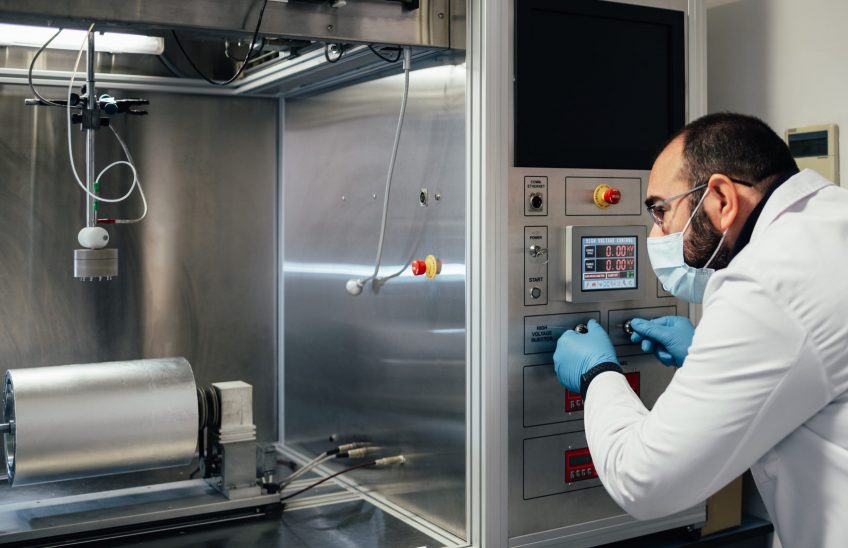
3. Making a conductive semi-organic polymer blend using electrospinning
Nanofibers comprise different materials and can be produced in a myriad of ways, such as drawing, electrospinning, self-assembly, template synthesis, and thermal-induced phase separation. Electrospinning is currently the most widely used method for the preparation of nanofibers. Electrospinning is a nanofiber production procedure. According to Alexander, “what you need for it is a syringe pump, a suitable polymer or polymer blend, a collector and a high voltage supply. So, you take your syringe pump with your polymer solution inside of it, you inject the polymer at a certain rate through a needle, you connect the needle tip to a high voltage power supply and the collector base is connected to ground potential.” Next, a polymer blend was necessary to produce the nanofibers. Alexander’s approach entailed “making a custom polymer blend using an organic semiconducting polymer”. He also implemented some secondary doping “which is done to improve the conductivity of your polymer”.
4. Future: Creating a nanofiber-based biosensor for the detection of targeted antigens
Commercially available antigen capture enzyme-linked immunosorbent assays (ELISAs) are used to analyse blood, organ suspension, plasma, or serum samples. For the final tests, a version of the COVID-19 South African variant, spike (S) glycoproteins, will be targeted which promote entry into cells and are the main target of antibodies. The initial focus of this research will be on resistance sensing to keep the research cost-effective for point of care purposes.
Read our recent articles:
Resistive Biosensor for the Detection of LC3 Protein in Autophagy
Participatory Research for Low-resourced Machine Translation: A Case Study in African Languages